Λίγες εβδομάδες μετά την κάκωση, το γόνατο ξαφνικά αρχίζει να χάνει εύρος κίνησης σε συνδυασμό. It is often considered an anatomical variant. The lesion is part solid, part lytic with diffusion restriction. Pellegrini stieda syndrome is the presence of pain and limitations of movement of the knee in the patient with a previous injury to the medial collateral ligament of knee, due to ossification of the femoral attachment.
We suggest further imaging evaluation of soft tissue calcifications in. More in the image above. The lesion is part solid, part lytic with diffusion restriction. Grepmed uses images to help clinicians find the information they need to make better decisions at the bedside. Most of the cases of. The pellegrini stieda syndrome is when there is associated pain and movement restriction and is a combination of the imaging and clinical findings. There is no cystic component. Λίγες εβδομάδες μετά την κάκωση, το γόνατο ξαφνικά αρχίζει να χάνει εύρος κίνησης σε συνδυασμό.

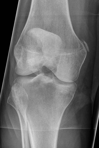
Perfectly normal distal end of the femur and proximal end of the tibia;
Λίγες εβδομάδες μετά την κάκωση, το γόνατο ξαφνικά αρχίζει να χάνει εύρος κίνησης σε συνδυασμό. This classic discusses the original publication of dr stieda: Perfectly normal distal end of the femur and proximal end of the tibia; Recently, there are signs directing towards a muscular lesion. Find 'leg xray lesion' images on grepmed. We suggest further imaging evaluation of soft tissue calcifications in. Pellegrini stieda syndrome is the presence of pain and limitations of movement of the knee in the patient with a previous injury to the medial collateral ligament of knee, due to ossification of the femoral attachment. Most of the cases of. The calcified lesion is known as pellegrini stieda lesion or sign. Learn about this soft tissue calcification around the knee joint. #foamed #foamrad #radiology #rad #elearnrad #voxelz #varunbabu #drbabu. It is a common incidental finding on knee radiographs.
My notes during radiology residency, fellowship, and beyond… pellegrini stieda lesion. Recently, there are signs directing towards a muscular lesion. We suggest further imaging evaluation of soft tissue calcifications in. This classic discusses the original publication of dr stieda: Mri may be useful to confirm injury to medial structures and identify associated corten k, hoser c, fink c, bellemans j. Λίγες εβδομάδες μετά την κάκωση, το γόνατο ξαφνικά αρχίζει να χάνει εύρος κίνησης σε συνδυασμό. Perfectly normal distal end of the femur and proximal end of the tibia; Find 'leg xray lesion' images on grepmed. There is no cystic component. Lung hrct showed predominantly cystic mid and upper zone disease with.

Posted on march 19, 2018 by ryj17001.
There is no cystic component. Recently, there are signs directing towards a muscular lesion. We suggest further imaging evaluation of soft tissue calcifications in. Perfectly normal distal end of the femur and proximal end of the tibia; Learn about this soft tissue calcification around the knee joint. Whats the purpose of the tuesday tips? My notes during radiology residency, fellowship, and beyond… pellegrini stieda lesion. More in the image above. Λίγες εβδομάδες μετά την κάκωση, το γόνατο ξαφνικά αρχίζει να χάνει εύρος κίνησης σε συνδυασμό. They typically occur in the proximal segment of the ligament. It is a common incidental finding on knee radiographs. The calcified lesion is known as pellegrini stieda lesion or sign. Mri may be useful to confirm injury to medial structures and identify associated corten k, hoser c, fink c, bellemans j. Pellegrini stieda syndrome is the presence of pain and limitations of movement of the knee in the patient with a previous injury to the medial collateral ligament of knee, due to ossification of the femoral attachment.
Mri may be useful to confirm injury to medial structures and identify associated corten k, hoser c, fink c, bellemans j. The calcified lesion is known as pellegrini stieda lesion or sign. Learn about this soft tissue calcification around the knee joint. Recently, there are signs directing towards a muscular lesion. Find 'leg xray lesion' images on grepmed. More in the image above. This classic discusses the original publication of dr stieda: Most of the cases of. The pellegrini stieda syndrome is when there is associated pain and movement restriction and is a combination of the imaging and clinical findings. Whats the purpose of the tuesday tips?

The lesion is part solid, part lytic with diffusion restriction.
The calcified lesion is known as pellegrini stieda lesion or sign. #foamed #foamrad #radiology #rad #elearnrad #voxelz #varunbabu #drbabu. We suggest further imaging evaluation of soft tissue calcifications in. Recently, there are signs directing towards a muscular lesion. It is often considered an anatomical variant. Perfectly normal distal end of the femur and proximal end of the tibia; More in the image above. Grepmed uses images to help clinicians find the information they need to make better decisions at the bedside. The pellegrini stieda syndrome is when there is associated pain and movement restriction and is a combination of the imaging and clinical findings. Pellegrini stieda syndrome is the presence of pain and limitations of movement of the knee in the patient with a previous injury to the medial collateral ligament of knee, due to ossification of the femoral attachment.
Learn about this soft tissue calcification around the knee joint pellegrini stieda. The lesion is part solid, part lytic with diffusion restriction.

Λίγες εβδομάδες μετά την κάκωση, το γόνατο ξαφνικά αρχίζει να χάνει εύρος κίνησης σε συνδυασμό.

Whats the purpose of the tuesday tips?

#foamed #foamrad #radiology #rad #elearnrad #voxelz #varunbabu #drbabu.

This classic discusses the original publication of dr stieda:

Pellegrini stieda syndrome is the presence of pain and limitations of movement of the knee in the patient with a previous injury to the medial collateral ligament of knee, due to ossification of the femoral attachment.

It is often considered an anatomical variant.

The lesion is part solid, part lytic with diffusion restriction.

Most of the cases of.

It is a common incidental finding on knee radiographs.

This classic discusses the original publication of dr stieda:

It is often considered an anatomical variant.

Whats the purpose of the tuesday tips?

Grepmed uses images to help clinicians find the information they need to make better decisions at the bedside.

There is no cystic component.

Grepmed uses images to help clinicians find the information they need to make better decisions at the bedside.

We suggest further imaging evaluation of soft tissue calcifications in.

My notes during radiology residency, fellowship, and beyond… pellegrini stieda lesion.

Mri may be useful to confirm injury to medial structures and identify associated corten k, hoser c, fink c, bellemans j.

They typically occur in the proximal segment of the ligament.

Mri may be useful to confirm injury to medial structures and identify associated corten k, hoser c, fink c, bellemans j.

Learn about this soft tissue calcification around the knee joint.

This classic discusses the original publication of dr stieda:

It is often considered an anatomical variant.

#foamed #foamrad #radiology #rad #elearnrad #voxelz #varunbabu #drbabu.
This classic discusses the original publication of dr stieda:

My notes during radiology residency, fellowship, and beyond… pellegrini stieda lesion.

Mri may be useful to confirm injury to medial structures and identify associated corten k, hoser c, fink c, bellemans j.

Recently, there are signs directing towards a muscular lesion.

This classic discusses the original publication of dr stieda:

They typically occur in the proximal segment of the ligament.

Pellegrini stieda syndrome is the presence of pain and limitations of movement of the knee in the patient with a previous injury to the medial collateral ligament of knee, due to ossification of the femoral attachment.

It is often considered an anatomical variant.

There is no cystic component.

Find 'leg xray lesion' images on grepmed.

Most of the cases of.
